![]()
University Hospital Basel

National Centre of Competence in Nanoscale Science, University of Basel
Polymer nanocontainers for receptor-specific cell targeting |
What is a nanocontainer? |
|
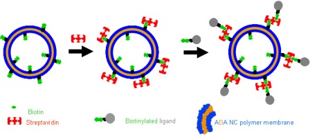
Preparation of nanocontainers for receptor targeting: NC’s containing 10% biotinylated copolymer were incubated with streptavidin solution, were separated chromatographically, and were incubated with the biotinylated ligand. |
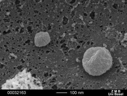
Scanning electron microscopy (SEM) micrograph of nanocontainers shows roughly spherical objects with sizes ranging from 100-250nm diameter. Note that the drying step necessary for SEM might somewhat alter the spherical appearance of nanocontainers as seen in reality.
|
What is the background of this nanocontainer project? |
What are the main results of this nanocontainer project? |
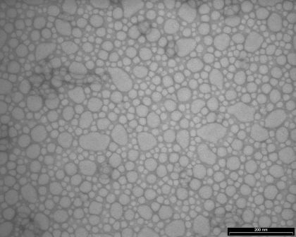
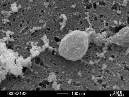
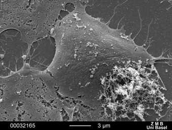
Polymer nanocontainers seen by transmission (left) and scanning electron microscopy (right)
|
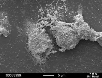
Main cell types we are working with are human THP-1 macrophages (left) and simian COS-7 cells
|
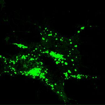
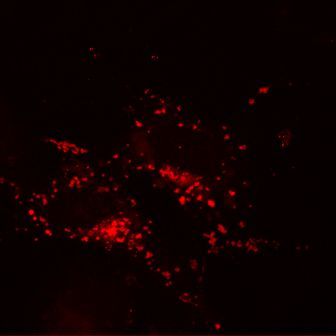
Transgenic COS-7 cells expressing the target receptor scavenger receptor A1 (SRA-1) were treated with nanocontainers coupled to the tracking ligand PolyG. Analysis was done by confocal microscopy (FITC filter set left, Texas Red filter set right). The left picture shows the localization of the cellular SRA-1 receptor, which were labeled by co-expression of enhanced green fluorescent protein (EGFP). The right picture shows the localization of the nanocontainers, which were filled with a red fluorescent dye. The pictures demonstrate the receptor-ligand co-localization, an important condition for receptor-selective targeting of nanocontainers. Furthermore, it is evident that the target cell stores the endocytosed nanocontainers in intracellular vesicles - most probably lysosomes. (click here for further details)
|
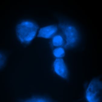
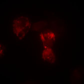
Scavenger receptor A1-expressing macrophages derived from human THP-1 cells were treated with fluorescent, ligand-labeled nanocontainers. Analysis by fluorscent microscopy (Dapi filter left, Texas Red filter right) shows the localization of the cells by weak autofluorescence (left). The localization of the nanocontainers is shown by the red fluorescent content (right). The cytosol of the cell is filled with vesicular accumulations of fluorescence, while the cell nucleus shows no signal. |